
What Is Retinal Vein Occlusion (RVO)?
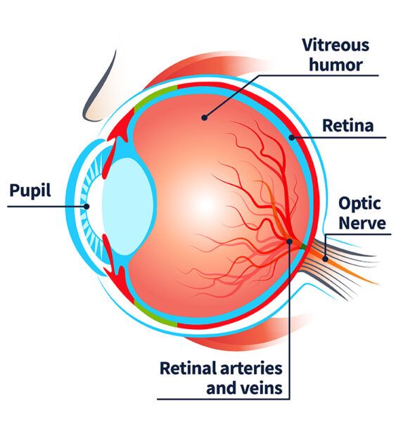
Retinal vein occlusion (RVO) is the partial or total blockage of a vein that drains blood from the retina. The retina, located in the back of the eye, captures images and sends them to the brain through the optic nerve to help us see. This blockage of the retinal vein can lead to vision loss and blindness. RVO is the second most common disease in the world that affects blood vessels in the retina, following diabetic retinopathy.
What Causes Retinal Vein Occlusion?
RVO occurs when blood flow is blocked in a vein in the retina by:
- Compression of a retinal vein by a retinal artery
- Slowdown of blood flow due to increased eye pressure
- A blood clot
Am I At-Risk for RVO?
There are several factors that can increase your risk for RVO, including:
- Age, over 40
- High blood pressure
- Atherosclerosis (ath-uh-roh-skluh-roh-sis) – hardening of the arteries due to plaque build-up
- Diabetes
- Hyperlipidemia (hy-per-lip-id-emia) – high levels of lipids (fats) in the blood
- Glaucoma
- History of RVO in your other eye
- Cigarette smoking
- Kidney disease
- Certain blood clotting disorders
What are the Different Types of Retinal Vein Occlusion
There are two main types of RVO, based on the location of where normal blood flow is being blocked.
Central Retinal Vein Occlusion (CRVO): the main vein in your eye is blocked
Branch Retinal Vein Occlusion (BRVO): small blood vessels attached to the main vein in your eye are blocked
What Are the Symptoms of Retinal Vein Occlusion?
How Does RVO Affect My Eyes?
RVO affects the eyes in several ways, and can often lead to vision loss and blindness if left untreated.
Retinal swelling (macular edema): swelling in the center of your retina that may cause blurry vision or loss of vision.
Lack of blood flow (ischemia): areas of the retina lose proper blood flow, and this can lead to vision loss.
Abnormal blood vessel formation (neovascularization of the eye): new blood vessels may form in the retina or iris.
Bleeding inside the eye (vitreous hemorrhage): caused by the formation of abnormal blood vessels in the eye that often break and leak; can lead to sudden, but painless, vision loss in that eye.
Increased eye pressure (neovascular glaucoma): eye pressure may reach dangerous levels as a result of abnormal blood vessel formation in the eye.
Retinal detachment: abnormal retinal blood vessels may cause the retina to pull away from its regular position, which interrupts oxygen and nutrient flow in the eye.

Blurry Vision

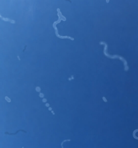
Floaters
How is Retinal Vein Occlusion Diagnosed?
Eye care providers diagnose RVO through an eye exam and retinal imaging tests. They can also work with your retina specialist and primary care doctor to determine the cause of retinal blood flow issues and lower your risk for future issues.
Eye Exam
The eye care provider will look closely at the outside and inside of your eye. To see inside, they will dilate your pupils to examine the back of your eye. This eye exam can help to:
- Determine which type of RVO you have
- Identify signs of swelling in the eye and abnormal blood vessel formation
- Estimate the lack of blood flow from your retina
- Plan your eye treatment
Retinal Imaging
The eye care provider may also take high-quality digital photos of the back of your eye, through a process called retinal imaging. This process helps to identify and monitor eye complications, including abnormal blood vessel formation, bleeding inside the eye, eye swelling, and retinal vein blockage
- Retinal photographs: photos taken of the inside surface of the eye.
- Fluorescein angiography: fluorescent dye is injected into the vein of the arm or hand, and pictures of the retina are taken through the dilated pupils.
- Optical Coherence Tomography (OCT): Cross-sectional pictures of the retina are taken to map and measure the thickness of the retina.
What Treatments Are Available for Retinal Vein Occlusion?
Currently, there is no treatment to reverse or cure retinal vein occlusion. However, there are several ways to prevent or treat complications that may be experienced because of the disease.
Anti-VEGF injections
Anti-VEGF injections are usually the first choice of treatment to help reduce swelling and leakage in the back of your eye. These injections help reduce the formation of abnormal blood vessels, which can leak and cause eye swelling.
Steroid injections
For patients who do not respond to anti-VEGF, steroid injections can help reduce eye swelling. Typically used as a second option following anti-VEGF injections because steroid injections may cause increased eye pressure and cataracts in some individuals. Note: long-term use of steroids may lead to complications. Talk to your doctor for more information.
Panretinal laser photocoagulation (PRP)
This laser procedure targets damaged areas of the retina to help prevent the formation of abnormal blood vessels and related bleeding in the eye. It can also help stabilize eye pressure.
Vitrectomy surgery (posterior pars plana vitrectomy (PPV))
This procedure helps to treat RVO patients with retinal detachment and/or severe, long-term bleeding that keeps coming back. The damaged vitreous humor, the gel-like fluid that fills the center of the eyeball, is removed and the damaged retina is repaired. PRP is typically given at the same time as well.
Can Vision Improve After RVO Treatment?
Yes, most of the patients treated for RVO experience significant improvement in vision. However, it is rare to get back 20/20 vision. Responses to treatment vary depending on how severe the disease is, the extent of damage to the retina, and the length of time with the disease before starting treatment. It is important to start treatment early for better outcomes.
What Can I Do to Prevent RVO?
Manage health conditions
Talk to your doctor to see if medications can help manage other related health conditions, such as high blood pressure, diabetes, or high cholesterol.
Exercise
Staying active as part of a healthy lifestyle can reduce your risk of developing problems that can lead to eye disease, and can improve your overall health.
Avoid smoking and tobacco products
Smoking increases the risk of eye damage. Quit smoking to help not only your eyes, but your overall health.
Schedule a routine eye exam
Your eye care provider can detect conditions at a routine eye exam, which can lead to earlier treatment and better outcomes.
Related Resources






This resource is funded with support from:

Prevent Blindness retains full independence over all our health education content, policy positions, advocacy efforts, and programmatic decisions. Find out more about Prevent Blindness and corporate sponsorships.